 Skip to content
As of 2025, CRISPR-Cas9 genome editing has transitioned from research tool to clinical application, with FDA-approved therapies such as Casgevy (exagamglogene autotemcel) for sickle cell disease and transfusion-dependent β-thalassemia. This clinical adoption has intensified demand for robust, reproducible laboratory workflows in both academic and industrial settings.
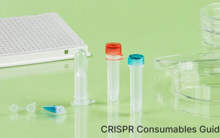
This guide provides a comprehensive catalog of laboratory consumables required across the CRISPR experimental workflow—from gRNA synthesis and delivery to clonal isolation. For each consumable category, we specify the technical parameters that directly influence experimental outcomes, including RNase/DNase-free certification, endotoxin limits, protein adsorption characteristics, and material compatibility requirements. The objective is to serve as a reference for procurement specifications and quality control criteria in CRISPR-based genome editing experiments.
What Is a CRISPR Experiment?
CRISPR-Cas9 genome editing enables targeted modification of DNA sequences through a ribonucleoprotein complex composed of the Cas9 endonuclease and a guide RNA (gRNA). The gRNA directs Cas9 to a specific genomic locus via complementary base pairing, where Cas9 introduces a double-strand break. Cellular repair mechanisms—non-homologous end joining (NHEJ) or homology-directed repair (HDR)—then generate insertions, deletions, or precise sequence replacements.
From a consumables perspective, two biochemical properties dictate material requirements:
(1) gRNA is highly susceptible to degradation by RNase contamination, necessitating RNase-free plasticware throughout synthesis and handling;
(2) Cas9 protein exhibits non-specific adsorption to standard polystyrene surfaces, requiring low-binding consumables during RNP complex assembly and delivery. These characteristics establish baseline quality criteria for laboratory supplies used in CRISPR workflows.
gRNA Preparation
This stage involves the synthesis and purification of guide RNA (gRNA), either through chemical synthesis or in vitro transcription (IVT) from a DNA template. The objective is to obtain intact, sequence-accurate gRNA free of enzymatic contaminants that could compromise downstream editing efficiency.
Key consumables and specifications:
RNase-free microcentrifuge tubes (0.2 mL, 1.5 mL, 2.0 mL)
Must be certified RNase-free and DNase-free, with independent sterility validation per batch. Packaging should maintain integrity during storage at –20 °C to –80 °C.
Filter tips recommended for gRNA handling to prevent aerosol contamination. Material must not leach RNase under standard laboratory conditions.
Low-binding PCR tubes (0.2 mL)
Required for IVT reactions to minimize adsorption of RNA polymerase and nascent RNA transcripts. Surface treatment should reduce protein binding by ≥90 % compared to standard polystyrene.

Silica-membrane columns must retain structural integrity during centrifugation (≤8,000 × g). Eluted RNA should yield A260/A280 ratios between 1.9 and 2.1, indicating minimal protein or solvent contamination.

For long-term gRNA storage at –80 °C. Tubes must feature O-ring seals rated for temperatures down to –196 °C and withstand ≥50 freeze-thaw cycles without leakage or structural failure.

All consumables contacting gRNA prior to RNP complex formation must be validated for RNase absence using a sensitive detection method (e.g., fluorescence-based assay with detection limit ≤0.01 ng/μL).
Plasmid Construction and Purification
This stage encompasses restriction enzyme digestion, ligation of gRNA sequences into expression vectors, bacterial transformation, and subsequent plasmid isolation. The objective is to generate endotoxin-free, high-purity plasmid DNA suitable for mammalian cell transfection.
Key consumables and specifications:
Silica-based adsorption columns must yield plasmid DNA with endotoxin levels ≤0.1 EU/μg for transfection-grade applications. A260/A230 ratios should exceed 2.0, indicating minimal salt or solvent carryover.
Low-binding microcentrifuge tubes (1.5 mL)
Required for enzymatic reactions (digestion, ligation) to minimize adsorption of Cas9 protein or restriction enzymes. Protein binding should be ≤5 % relative to standard polystyrene under reaction conditions.
Bacterial culture consumables
Sterile LB broth bottles and agar plates must support consistent colony formation. Plates should maintain moisture retention for ≥48 h at 37 °C without condensation interference.
Transformation tubes
Thin-walled microcentrifuge tubes for chemical transformation must withstand 42 °C heat shock without deformation. Pre-chilling to 0–4 °C prior to use is required to maintain competent cell viability.
Sterile inoculation loops and spreaders
Disposable plastic loops must be certified DNA-free to prevent cross-contamination between clones during colony picking.
All plasticware contacting plasmid DNA prior to transfection must be certified endotoxin-free and DNase-free. For transfection applications, plasmid preparations should be quantified by fluorometric methods (e.g., Qubit) rather than UV absorbance alone to avoid RNA contamination artifacts.
Cell Transfection and RNP Delivery
This stage delivers CRISPR components into target cells via plasmid transfection or direct ribonucleoprotein (RNP) electroporation. The objective is to achieve efficient intracellular delivery while maintaining cell viability above 70 % at 24 h post-treatment.
Key consumables and specifications:
Transfection-grade microcentrifuge tubes
Low-binding tubes required for complex formation (e.g., lipid–DNA or RNP assembly). Must be certified endotoxin-free (<0.1 EU/mL) and compatible with serum-free media during complex incubation.

Serum-free media
Required during lipid-based transfection complex formation. Must support complex stability for 15–20 min without precipitation. Standard culture media containing serum should not be used during this step.
Electroporation cuvettes
For RNP delivery: 2 mm or 4 mm gap cuvettes depending on cell type. Must be single-use to prevent arcing from residual salts or cellular debris. Material must withstand field strengths up to 1,500 V/cm without dielectric breakdown.

Tissue-culture treated polystyrene dishes (35–100 mm), multi-well plates (6–96 wells), and flasks (T25–T175) must provide consistent surface hydrophilicity (contact angle 40–70°) to support uniform cell attachment. Batch-to-batch variation in cell adhesion should not exceed ±15 %.

Cryogenic vials for cell banking
External-thread vials with silicone gaskets required for post-editing cell storage. Must maintain seal integrity after immersion in liquid nitrogen (–196 °C) and withstand ≥30 freeze-thaw cycles without leakage.
All consumables contacting live cells must be sterile (SAL 10⁻⁶) and free of cytotoxic leachables as validated by ISO 10993-5 extractable testing. For electroporation, cuvettes must be stored desiccated and brought to room temperature prior to use to prevent condensation-induced arcing.
Editing Efficiency Assessment
This stage evaluates the frequency of on-target modifications using enzymatic mismatch cleavage, sequencing, or protein-level detection methods. The objective is to quantify editing efficiency and identify unintended indels or off-target effects.
Key consumables and specifications:
PCR tubes for amplification
Low-binding 0.2 mL tubes required for target locus amplification prior to T7E1 assay or sequencing library preparation. Tubes must not inhibit high-fidelity DNA polymerases at standard cycling conditions.
T7 Endonuclease I assay reagents
Microcentrifuge tubes for heteroduplex digestion must be certified nuclease-free. Reaction vessels should maintain temperature stability during 37 °C incubation without condensation on inner walls.
Agarose gel electrophoresis supplies
Gel trays and combs must be chemically resistant to TAE/TBE buffers. DNA ladders and loading dyes should be stored in RNase/DNase-free tubes to prevent degradation during repeated use.
Sanger sequencing purification columns
Required for PCR product cleanup prior to capillary sequencing. Eluted DNA must yield A260/A230 ratios >1.8 to avoid signal suppression during electrophoresis.
NGS library preparation consumables
Magnetic beads for size selection must exhibit batch-consistent binding capacity (CV <10 %). Low-binding 96-well plates required for library quantification by qPCR; plates must be optically clear with minimal autofluorescence at 488 nm excitation.
Flow cytometry tubes
12 × 75 mm polystyrene tubes for reporter-based assays must be free of static charge to prevent cell adhesion to tube walls. Tube geometry must be compatible with standard flow cytometer sample injectors.
All consumables used in detection workflows must be free of DNA/RNA contaminants that could generate false-positive signals. For NGS applications, library quantification should be performed using fluorometric methods to avoid overestimation from adapter dimers or primer artifacts.
Clonal Isolation
This stage isolates single edited cells and expands them into monoclonal populations for genotypic validation. The objective is to obtain pure clonal lines with defined editing outcomes (homozygous, heterozygous, or wild-type) while minimizing cross-contamination and maintaining cell viability during expansion.
Key consumables and specifications:
Required for limiting dilution to concentrate single cells at the well center. Bottom curvature radius should be 2.0–3.0 mm. Plates must maintain sterility for ≥14 days under standard incubation conditions.
384-well plates for FACS deposition
Used for single-cell sorting via fluorescence-activated cell sorting. Wells must accommodate ≤1 μL droplet volumes with minimal evaporation (<5 % over 72 h at 37 °C, 5 % CO₂). Optical clarity required for post-sort imaging.

Low-evaporation sealing films
Adhesive films or breathable seals must reduce edge-well evaporation to <10 % over 96 h. Material should permit gas exchange while preventing contamination.
Cloning cylinders and scrapers
Sterile stainless-steel or disposable plastic rings for manual clone picking. Inner diameter typically 5–8 mm. Must be certified DNA-free to prevent cross-contamination between clones.
Genotyping PCR plates
Low-binding 96-well plates for colony PCR. Must be compatible with direct lysis protocols and exhibit minimal inhibition of Taq polymerase. Optical grade required if real-time PCR is used for genotyping.
Cryopreservation vials for clone banking
External-thread vials with silicone gaskets for long-term storage of validated clones. Must maintain seal integrity after liquid nitrogen immersion and withstand ≥20 freeze-thaw cycles.
All consumables used during clonal expansion must be sterile and free of leachable compounds that impair cell proliferation. For FACS-based isolation, plate geometry must comply with instrument manufacturer specifications to ensure accurate single-cell deposition.
Conclusion
CRISPR genome editing relies on a defined set of laboratory consumables across five workflow stages: gRNA preparation, plasmid construction, cell delivery, editing validation, and clonal isolation. Each stage imposes specific material requirements—including RNase-free certification for RNA handling, low-binding surfaces for protein preservation, endotoxin limits <0.1 EU/μg for transfection, and evaporation control during clonal expansion. Adherence to these specifications supports reproducible editing outcomes and minimizes experimental variability.
For laboratories seeking consumables that meet these technical criteria, GenFollower provides a curated catalog of laboratory supplies validated for genome editing applications. All products undergo batch-specific quality testing against the parameters outlined in this guide. Visit genfollower.com to access specifications and procurement documentation.
Recent Posts
CRISPR Consumables Guide: The Complete Lab Supply List for gRNA Synthesis, Transfection & Clonal Screening
As of 2025, CRISPR-Cas9 genome editing has transitioned from research tool to clinical application, with FDA-approved therapies such as Casgevy (exagamglogene autotemcel) for sickle cell disease and transfusion-dependent β-thalassemia. This clinical adoption has intensified [...]

Common Techniques for Isolating Microorganisms on Solid Media
In the natural environment, microorganisms are seldom found in isolation; instead, they typically thrive within complex, multi-species consortia across diverse habitats, ranging from soil and aquatic ecosystems to the human gut. For researchers aiming to [...]
The Ultimate Guide to Agarose Gel Electrophoresis
Agarose gel electrophoresis is a standard technique used in molecular biology for the separation, identification, and purification of nucleic acids. Its operational simplicity and broad applicability make proficiency in this method a fundamental prerequisite for [...]
